How Are Fibroids Removed While Preserving the Uterus?
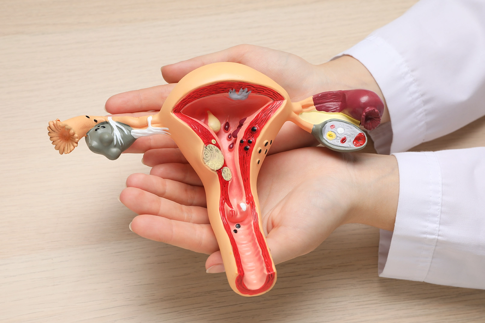

When fibroids require surgery, preserving the uterus may be an important consideration, particularly for women planning a future pregnancy. Yet if a fibroid is embedded within the uterine wall, how can it be removed without taking out the uterus itself?
Myomectomy allows selected fibroids to be removed while leaving the uterus in place. For women considering this procedure, understanding how it works can provide greater clarity about the proposed treatment and help guide discussions with their gynaecologist.
How Is Uterus-Preserving Fibroid Removal Planned?
Before choosing a surgical approach, the surgeon needs to understand how each fibroid relates to the uterine wall. This helps determine how the growth can be reached and how much surrounding tissue may be affected during removal.
Assessing Fibroid Size, Location and Appearance
A pelvic ultrasound is commonly used to assess the size, number and location of fibroids. An MRI scan may provide further detail when fibroids are numerous or deeply embedded in the uterine muscle. These findings help the surgeon plan how the fibroids can be accessed and removed.
The location of each fibroid is particularly important:
- Within the uterine wall (intramural): These fibroids are surrounded by muscle and may require an incision into the uterine tissue.
- Along the outer surface (subserosal): These may be easier to access, depending on how deeply they extend into the muscle.
- Beneath the inner lining (submucosal): These protrude towards or into the uterine cavity and may be removable through the cervix.
Fibroids that distort the uterine cavity may also affect fertility, making their relationship to the uterine lining another important consideration during surgical planning. Beyond their size and location, imaging also allows the surgeon to assess the appearance of the fibroids and identify any unusual features that warrant further evaluation before surgery.
Choosing the Surgical Approach
Once the surgeon has assessed the fibroids through imaging, the next step is to determine the most suitable way to remove them while preserving the uterus. This involves considering whether the fibroids can be accessed through the cervix or require abdominal surgery.
- Laparoscopic myomectomy: May be suitable when fibroids can be accessed and removed through small abdominal incisions, with enough room for the surgeon to repair the uterine wall where necessary.
- Open myomectomy: May be preferred for particularly large or numerous fibroids, especially when wider access is needed to remove them and manage the surrounding uterine tissue.
- Hysteroscopic myomectomy: Generally suitable for fibroids that protrude into the uterine cavity and are not too deeply embedded in the muscular wall. Instruments are passed through the vagina and cervix, avoiding abdominal incisions.
What Happens During a Myomectomy Procedure?
Although the method of accessing the fibroids varies according to the surgical approach, a myomectomy procedure generally involves identifying the growths, separating them from surrounding tissue and removing them while controlling bleeding.
Accessing and Separating the Fibroids
The surgeon first accesses the fibroids using the planned surgical approach. For laparoscopic or open myomectomy, this involves reaching the uterus through abdominal incisions. Fibroids embedded within the uterine muscle generally require an incision in the uterine wall to expose them.
Once exposed, the surgeon carefully separates each fibroid from the surrounding muscle, following the boundary between the growth and healthy tissue. Fibroids often have a distinguishable boundary from the surrounding muscle. By gently following this boundary, the surgeon can gradually separate the growth without unnecessarily cutting into healthy tissue. Particular care is taken when a fibroid lies close to the uterine cavity to minimise injury to its inner lining.
For hysteroscopic myomectomy, instruments are passed through the cervix to reach fibroids protruding into the uterine cavity. These fibroids are removed from within the cavity rather than through an abdominal incision.
Removing Fibroid Tissue and Controlling Bleeding
After separating the fibroids from the uterine tissue, the surgeon removes them using a method appropriate for their size and the surgical approach. Fibroids may be extracted intact or divided into smaller pieces when necessary. Tissue division, known as morcellation, carries specific risks, including the potential spread of unsuspected abnormal tissue, so the extraction method and precautions are considered during surgical planning.
Bleeding is also managed throughout the procedure, as fibroids and the surrounding uterine tissue contain blood vessels. The surgeon may use medication to reduce blood flow temporarily or surgical instruments to control bleeding from affected vessels.
Once the fibroids have been removed, the surgeon checks the treated areas for bleeding and assesses the uterine tissue before proceeding with any necessary repair. Removed tissue is typically sent for laboratory examination.
How Is the Uterus Repaired After Fibroid Removal?
The extent of uterine repair depends on how deeply the fibroids were embedded and how much muscle was opened during removal. Even when surgery is performed through small abdominal incisions, the uterine wall may require careful reconstruction to restore its structural integrity.
Closing Incisions in the Uterine Muscle
When removing a fibroid leaves an opening in the uterine muscle, the surgeon closes it using stitches. Deeper openings may require several layers of sutures to bring the muscle together and prevent spaces where blood could collect.
This repair can be performed during either laparoscopic or open myomectomy, using the surgical access already established for fibroid removal.
Hysteroscopic myomectomy generally does not require stitches because the fibroid is removed from within the uterine cavity without making the same type of incision through the uterine wall.
Allowing the Uterus to Heal
After laparoscopic or open myomectomy, the uterine muscle continues healing even after the abdominal wounds have closed, especially when deeper incisions were needed. Hysteroscopic myomectomy generally involves healing of the treated surface inside the uterine cavity, where scar tissue can sometimes form.
For women considering pregnancy, the depth of any uterine incisions may influence how long to wait before attempting conception and whether a caesarean delivery is recommended. Although myomectomy preserves the possibility of pregnancy, it does not guarantee fertility.
Discuss Uterus-Preserving Fibroid Surgery With Wei-Wei Clinic
If you are considering myomectomy surgery, a specialist consultation can help address your concerns by explaining whether the procedure is suitable for you, what the surgery involves and what to expect during recovery. Having a clearer understanding of the proposed treatment can provide reassurance and help you approach surgery with greater confidence.
At The Wei-Wei Clinic for Women, Senior Consultant Obstetrician & Gynaecologist Dr Wei-Wei Wee-Stekly assesses fibroid characteristics and individual treatment goals to determine whether myomectomy is suitable and which surgical approach may be appropriate. Dr Wei-Wei's surgical expertise allows her to tailor her surgical approach to each patient's condition, with an emphasis on careful tissue handling, uterine preservation and individualised care throughout treatment.
Book an appointment to discuss your fibroid treatment options and whether uterus-preserving surgery may be suitable for you.
Frequently Asked Questions (FAQs)
Meet Our Doctor
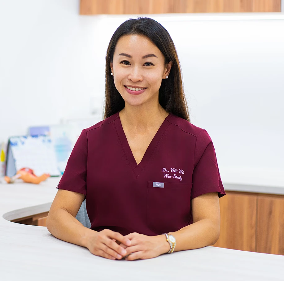
Dr Wei-Wei Wee-Stekly is a senior consultant obstetrician and gynaecologist with extensive experience in women's health at every stage of life. She earned her Bachelor of Medicine and Bachelor of Surgery from Guy's, King's and St Thomas' School of Medicine at King's College London in 2003. Dr Wee-Stekly went on to achieve her MRCOG in the United Kingdom and a Master of Medicine in Obstetrics and Gynaecology in Singapore. She is a Fellow of the Royal College of Obstetricians and Gynaecologists in the United Kingdom and a Fellow of the Academy of Medicine, Singapore.
More About Dr Wei-WeiWomen’s health and well-being are our priority.
For more information or to make an appointment, please call (65) 6250 5608 or leave us a message below.

Operating Hours:
Saturday: 08:30am – 12:30pm
Sunday and Public Holiday: Closed